
Visual Evoked Potential (VEP) Test
A visual evoked potential (VEP) test is a non-invasive test that measures the electrical activity in your brain when you see visual stimuli, such as flashing lights or patterns. It can help diagnose or monitor conditions that affect your optic nerves or visual pathways, such as multiple sclerosis (MS), optic neuritis, glaucoma, or brain tumors.
During a VEP test, electrodes are placed on the scalp to detect electrical signals generated by the brain in response to visual stimuli. These stimuli can be in the form of light flashes or patterns. By analyzing the waveform patterns of the brain’s electrical activity, researchers and clinicians can gain insights into the integrity of the visual pathway, as well as identify potential abnormalities or dysfunctions.
How does a VEP test work?
A VEP test works by recording the time it takes for the visual stimuli to reach your brain and trigger a response. The test uses electrodes that are attached to your scalp with a sticky gel. The electrodes measure the electrical signals that are generated by your brain when you see the stimuli.
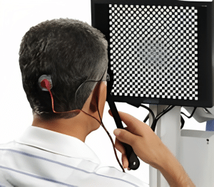
The stimuli are usually high-contrast patterns that flash and alternate on a screen in front of you. You will be asked to look at the center of the screen and keep your eyes open and still. Sometimes, a bright flashing light may be used instead of patterns, especially for children or people with impaired vision.
The test takes about 30 to 60 minutes to complete. You may be asked to do the test several times with different eyes or different types of stimuli. The test is painless and safe, but you may feel some discomfort from the electrodes or the flashing lights.
(Read More About Video EEG – Uses, Indications, Contraindications)
The Process of Visual Evoked Potential:
The process of conducting a Visual Evoked Potential test involves several steps:
- Preparation: Before the test, the patient’s scalp is prepared by cleaning it to ensure optimal electrode placement. Electrodes are then attached to specific areas of the scalp, typically following the International 10-20 system, which is a standardized method for electrode placement.
- Stimulus Presentation: Visual stimuli, such as checkerboard patterns or flashing lights, are presented to the patient. These stimuli are carefully designed to elicit specific responses from the visual system. The stimuli can be presented on a screen or through specialized goggles.
- Recording the Brain’s Response: As the patient views the visual stimuli, the electrodes detect the brain’s electrical activity in response to the stimuli. This activity is recorded and transformed into waveforms, which represent the brain’s response.
- Signal Analysis and Interpretation: The recorded waveforms are analyzed to identify specific components that correspond to different stages of visual processing. The latency, amplitude, and morphology of these components provide valuable information about the functioning of the visual system. Abnormalities or delays in the waveform patterns can indicate potential visual impairments or neurological disorders.
What can a Visual Evoked Potential test diagnose?
A VEP test can help diagnose or point to the following conditions:
- Multiple sclerosis (MS): MS is a chronic condition that causes your immune system to attack the myelin sheath that covers and protects your nerve fibers. This can cause inflammation and damage to your optic nerves and other parts of your central nervous system. A VEP test can detect delays or abnormalities in the transmission of visual signals from your eyes to your brain, which can indicate MS or optic neuritis (inflammation of the optic nerve). A VEP test is one of the tools that doctors use to diagnose MS, along with other tests such as MRI, spinal fluid analysis, blood tests, and neurological exams.
- Optic neuritis: Optic neuritis is a condition that causes inflammation and damage to your optic nerve, which carries visual information from your eye to your brain. It can cause symptoms such as blurred vision, pain in the eye, loss of color vision, or reduced peripheral vision. Optic neuritis can be caused by MS, infections, autoimmune disorders, or other factors. A VEP test can measure the extent of damage to your optic nerve and help determine the cause of optic neuritis.
- Glaucoma: Glaucoma is a group of eye diseases that damage your optic nerve due to high pressure inside your eye. It can lead to vision loss or blindness if left untreated. Glaucoma can affect your peripheral vision first, which may not be noticeable until it is advanced. A VEP test can detect changes in your peripheral vision and help diagnose glaucoma early.
- Brain tumors: Brain tumors are abnormal growths of cells in your brain or near your brain. They can be benign (non-cancerous) or malignant (cancerous). Brain tumors can affect your vision by pressing on your optic nerves or other parts of your visual system. A VEP test can help locate the tumor and assess its impact on your vision.
How are Visual Evoked Potential test results interpreted?
A VEP test produces a graph that shows the amplitude (height) and latency (time) of the electrical signals in your brain when you see the stimuli. The graph is called a waveform and it has peaks and valleys that correspond to different stages of processing visual information.
The most important peak is called P100 because it occurs around 100 milliseconds after seeing the stimulus. The latency of P100 reflects how fast the visual signal travels from your eye to your brain. The amplitude of P100 reflects how strong the signal is.
A normal P100 latency is between 80 and 120 milliseconds. A longer latency means that there is a delay in the transmission of visual signals, which can indicate damage or dysfunction in your optic nerves or visual pathways. A lower amplitude means that there is a reduction in the signal strength, which can indicate loss of nerve fibers or impaired function.
Advancements and Innovations in Visual Evoked Potential:
Over the years, technological advancements have enhanced the capabilities of Visual Evoked Potential testing. These advancements include:
- High-density Electrode Arrays: The use of high-density electrode arrays allows for more precise mapping of the visual system. With a greater number of electrodes, researchers can capture detailed information about the brain’s response to visual stimuli, leading to a more comprehensive understanding of visual processing.
- Advanced Signal Processing Techniques: Sophisticated signal processing techniques enable researchers to extract more precise and meaningful information from VEP waveforms. These techniques allow for better identification of subtle changes and abnormalities, improving the accuracy of diagnosis and research findings.
- Integration with Other Neuroimaging Techniques: Combining VEP with other neuroimaging techniques, such as functional magnetic resonance imaging (fMRI) or positron emission tomography (PET), allows for a more comprehensive investigation of the visual system. The integration of these techniques provides a multi-modal approach to understanding visual processing, enabling researchers to examine both the electrical and metabolic aspects of the brain’s response to visual stimuli.
Limitations and Future Directions:
While Visual Evoked Potential is a powerful tool, it does have limitations:
- Variability: VEP responses can vary among individuals, making it challenging to establish strict norms. Factors such as age, attention, and medication can influence the results. Therefore, careful interpretation and comparison to reference data are crucial.
- Limited Spatial Resolution: VEP provides information about the overall activity of the visual system but lacks precise spatial resolution. To overcome this limitation, researchers are exploring techniques such as high-density electrode arrays and integration with other imaging modalities.
As for future directions, ongoing research aims to refine VEP techniques, improve signal analysis methods, and explore novel applications. Additionally, the integration of VEP with emerging technologies like virtual reality and artificial intelligence holds promise for further advancements in understanding the visual system.
Your doctor will compare your VEP test results with normal values and with other tests to make a diagnosis or monitor your condition. Your doctor will also consider factors such as your age, eye condition, medication use, and other health issues that may affect your VEP test results.
FAQs
Is a Visual Evoked Potential test painful?
No, a VEP test is not painful. You may feel some discomfort from the electrodes or the flashing lights, but the test does not involve any needles, injections, or radiation.
Are there any risks or side effects of a Visual Evoked Potential test?
A VEP test is generally safe and has no serious risks or side effects. However, some people may experience headaches, nausea, dizziness, or eye strain from the flashing lights or patterns. If you have epilepsy or a history of seizures, you should inform your doctor before the test, as the flashing lights may trigger a seizure.
How should I prepare for a Visual Evoked Potential test?
You should follow your doctor’s instructions on how to prepare for a VEP test. You may be asked to avoid caffeine, alcohol, or certain medications that may affect your brain activity before the test. You should also wear comfortable clothing and glasses or contact lenses if you need them. You should not wear any makeup, hair products, or jewelry that may interfere with the electrodes.
How long does it take to get the results of a Visual Evoked Potential test?
It usually takes a few days to get the results of a VEP test. Your doctor will review your results and explain what they mean for your condition. You may need to have follow-up tests or appointments to confirm or monitor your diagnosis.
Takeaway
A VEP test is a useful tool that can help diagnose or monitor conditions that affect your vision or optic nerves, such as MS, optic neuritis, glaucoma, or brain tumors. It measures the electrical activity in your brain when you see visual stimuli and detects any delays or abnormalities in the transmission of visual signals. A VEP test is painless and safe, but you may experience some discomfort from the electrodes or the flashing lights. Your doctor will interpret your results and guide you on the next steps for your treatment or management.
References
(1) Visual Evoked Potential (VEP) Test for Multiple Sclerosis – Healthline.
(2) Evoked potentials | MS Trust.
(3) Evoked Potential Tests – What You Need to Know.
(4) Visually Evoked Potentials: Purpose, Preparation, Types – Verywell Health.
(5) Visual Evoked Potential (VEP Test): Procedure & Results – Cleveland Clinic.